RecommendMail Facebook LinkedIn
- Tech Talk
- Health & Science
How a microscope camera reveals secrets of marine microfossils for climate research
The use of a microscope camera in marine research helps in analyzing marine microfossils and reconstructing the environmental conditions in the course of the earth's climate history.
, Stefan SeidleinThe oceans are the regulating and stabilizing force of our climate system. They occupy approx. 70% of the earth's surface and 95% of the living biosphere. These figures alone reveal how important it is to understand the world's oceans and the enormous importance of polar and marine science for climate research as well as many other scientific disciplines.
Scientists at the Alfred Wegener Institute, Helmholtz Centre for Polar and Marine Research (AWI) explore the poles, the seas and the climate. We spoke with the marine researcher and micropaleontologist Dr Oliver Esper from the Geoscience Department to get an insight into his work. The research undertaken by this department looks back into the past by decoding data hidden in the ocean to reconstruct the climate history of our planet.
Dr Esper, what are you as marine researcher exploring, and what is the aim of your work as a micropaleontologist?
We analyze marine microfossils (remains of unicellular organisms) from the water column and the sediments both in the Micropaleontology Laboratories of the Geosciences Department at AWI and on sea-going expeditions. Our goal is to determine the species composition of fossil assemblages in order to reconstruct past environmental conditions, such as sea surface temperature, sea ice extent and the primary productivity of specific areas of the world ocean through the history of the earth.
Parts of the world’s oceans are key areas in influencing and steering changes in the global climate system. Marine sediments record these processes via changes in sedimentary processes and changes in the inventory of biological remains, such as fossil algae and tiny animal shells. The analysis of past changes in marine biology provides information on the amount and impact of oceanic influence on the climate system. Furthermore, marine sedimentary records of climate change are probably more complete (less eroded/altered) than terrestrial records, due to a more stable sedimentation environment and less disturbance of the sediment column.
What instruments and tools, such as a microscope camera do you need for your work as marine researcher?
I like to work with highly sophisticated equipment. In our AWI laboratories, we use several different types of microscopes. The JENOPTIK GRYPHAX® NAOS microscope camera is attached to microscopes to visualize and document siliceous, calcareous, and organic-walled hard parts of microorganisms gained from water samples, modern sea floor sediments, and ancient sediment core material primarily from the polar oceans. For 15 years in my work as a marine researcher, I have successfully used different models of Jenoptik microscope cameras on different microscopes.
Currently, we have chosen the JENOPTIK GRYPHAX® NAOS microscope camera as the new workhorse for our digital imaging because of its easy handling, high image quality and good price-performance ratio.
What is the procedure in marine research for this type of microscopic analysis?
First of all, the microfossils are removed from surrounding sediments or filtered from sea water, mounted on optical slides and then embedded with adhesive. Single objects or species assemblages are then analyzed, counted and documented at varying magnifications.
Digital images of single microfossils are archived to document specific findings and produce photo plates for scientific publications. The microscope camera JENOPTIK GRYPHAX® NAOS is therefore attached to different microscopes with specific analytical options, such as phase contrast and polarized light.
What objects do you specifically examine?
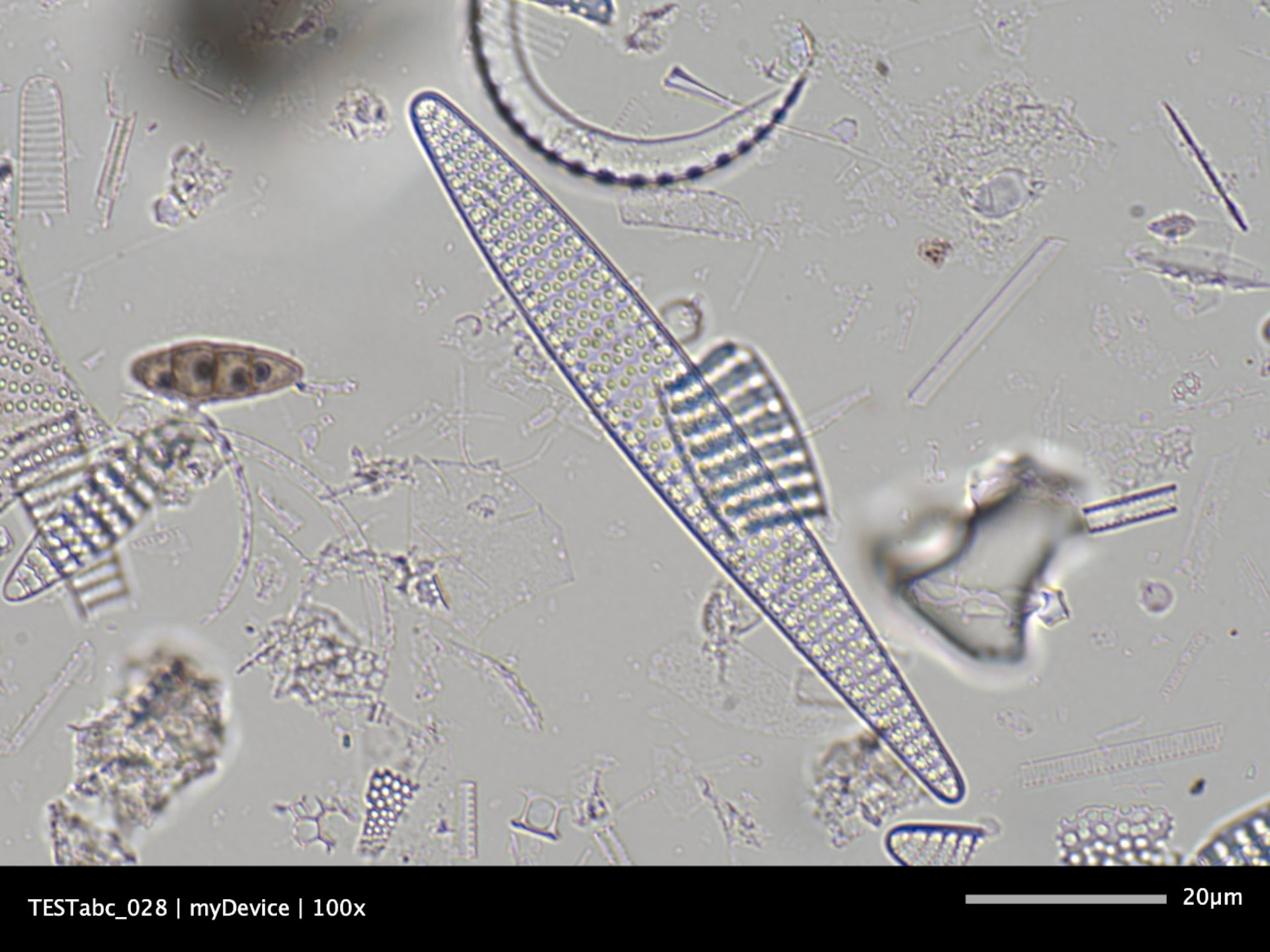
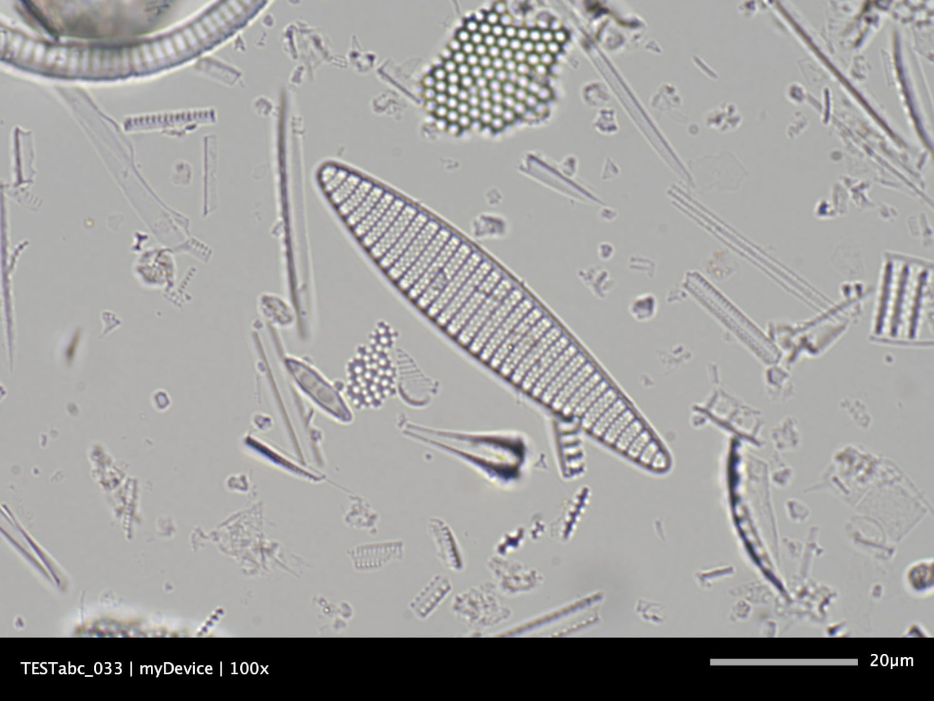
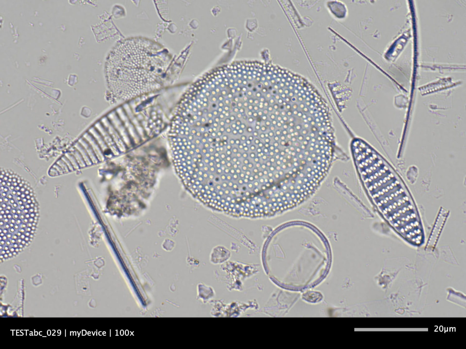
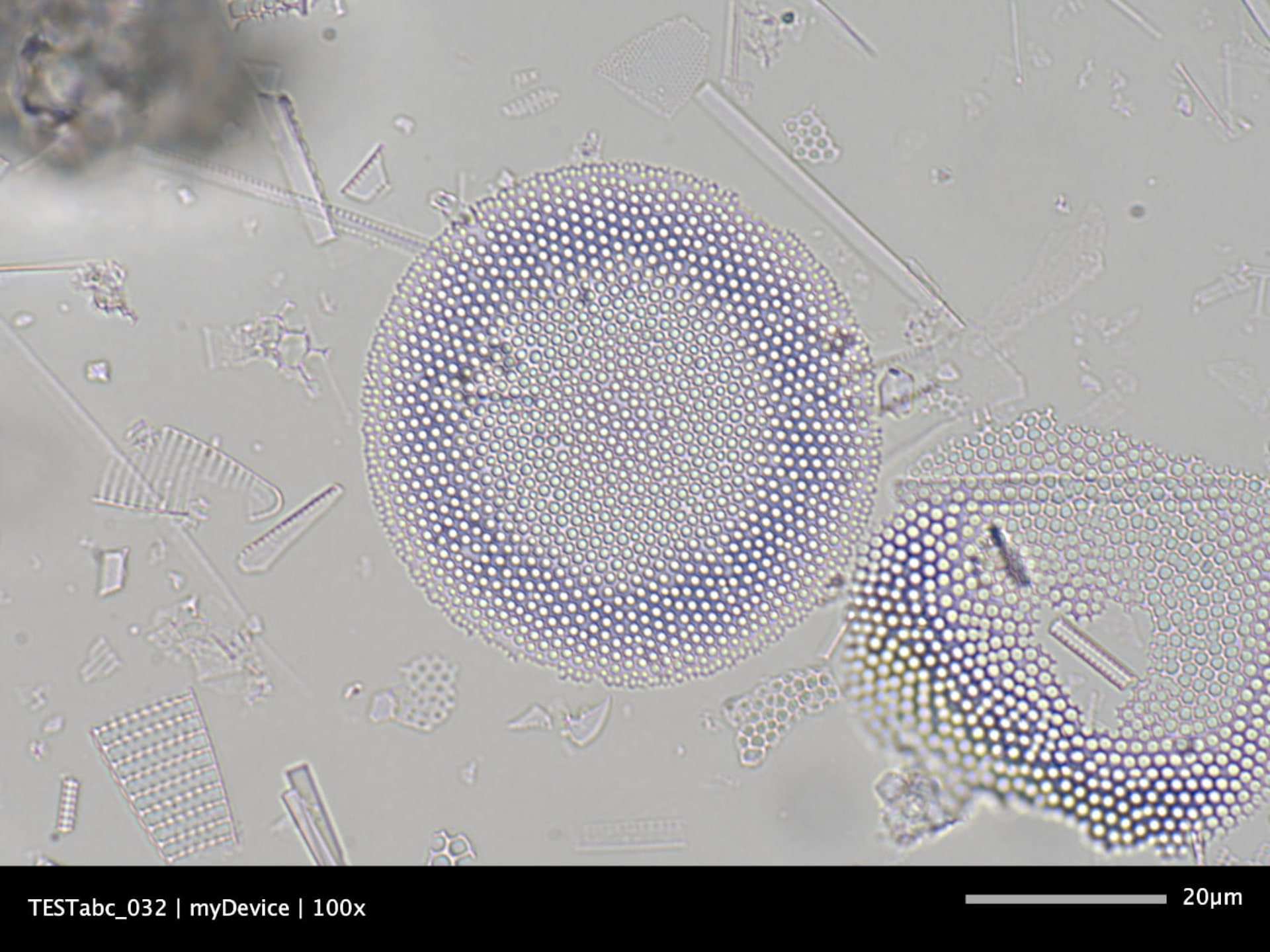
We see here 4 images, which I took with our microscope camera JENOPTIK GRYPHAX® NAOS and a lens with 100x magnification.
This is the dominant diatom species Fragilariopsis kerguelensis, which occurs in sediments of the Subantarctic.
The object on the top left is 90 microns in length and 10 microns in width, while the object on the top right has a length of 65 microns and a width of 12 microns.
In these two images below we see the diatom species Thalassiosira lentiginosa, which occurs abundantly in sediments of the Subantarctic.
The diatom on the bottom left, with a diameter of 45 microns, shows good preservation.
Are there any special requirements on the microscopic equipment that is used in marine research regarding the environmental conditions?
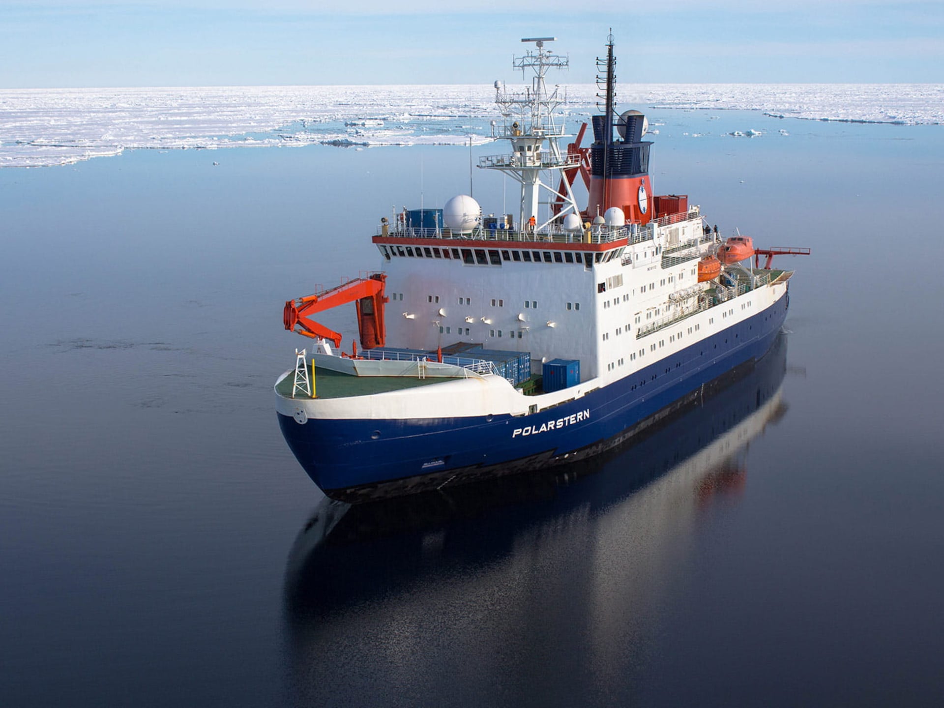
Absolutely, microscope workplaces can vary in nature – ranging from a clean, climate-controlled laboratory environment to ships’ laboratories with dusty, wet and salty air on research vessels, such as the RV POLARSTERN and RV SONNE. Over several months at sea in the remote areas of the Arctic and Antarctic oceans, the optical equipment has to meet special requirements.
The microscope camera has to be reliable and easy to operate to allow the immediate analysis of the samples on board. It also has to be robust in a climatically variable environment and must be able to produce good images – even under heavy ship movements and changing temperatures. The camera software has to be compatible with the ship's computer system, and image data needs to be stored easily.
This has been an exciting insight into the work of a marine researcher. Thank you very much Dr Esper.
Would you like to learn more about Jenoptik´s microscope cameras that play a significant role not only in geoscience and marine research but also in many other fields in healthcare & life science? Then do not hesitate to contact us.
The images are used with the kind permission of the Alfred Wegener Institute, Helmholtz Centre for Polar and Marine Research (AWI).Product recommendation

About Stefan Seidlein
Stefan Seidlein has been working for Jenoptik since 2000 in various positions in the field of Digital Imaging. As product manager, he currently focuses on the light microscope camera product portfolio and brings his entire digital imaging competence and experience to projects. As a graduated technician with a focus on energy technology and process automation, he is fascinated by digitalization and the many opportunities it offers both individuals and Jenoptik.