- Digitization
- Health & Science
- Tech Talk
How do doctors use optical technology to diagnose blood disorders? Hear it from an expert.
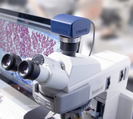
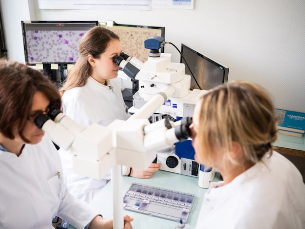
Precise hematological diagnosing requires a lot of experience and excellent true-color imaging from the microscope camera used for hematopathology – such as our GRYPHAX® ARKTUR camera. We interviewed an expert, Thomas Nebe, M.D., from the HaeMa laboratory in Mannheim, Germany to get a better understanding of the benefits of precise imaging for hematopathology diagnoses.
Dr. Nebe, in your opinion, what are the important aspects when using your JENOPTIK GRYPHAX® ARKTUR camera for hematological morphology?
In hematology, the smallest changes in color determine the diagnosis. The test specimens must be stained precisely so that even subtle changes can be seen. The staining method plays a very important role, with Wright staining and automated rapid staining being of limited value to hematopathologists. Reducing the intensity of staining does not show the structural details with adequate contrast. The gold standard for hematologists is the May-Grünwald-Giemsa staining according to Arthur von Pappenheim.
Other important aspects for hematologists are the true color reproduction, the contrast and the resolution of the camera used for the imaging. The hematologist diagnoses and records his findings based on the original microscope image as seen directly through the eyepiece. Those who send and receive the report, as well as others who may review it, can understand the report much better when it is based on what is written in the report combined with the microscopic image taken. A hematology camera should, therefore, offer not only high resolution but also very good high-contrast, true-color imaging in order to provide the data, especially that of microscopic image recording, as a reliable source for further evaluation by those who send, receive, and review it.
In addition, the true color imaging also increases productivity in the laboratory. The personnel costs of a conventional laboratory are approx. 75% of the overall costs for a sample submitted. The more quickly the hematologist analyzes the smear based on the provision of precise color information by the camera, the more efficient the entire laboratory process is, as there is no need to pick out or send the slide.
When asked why you chose a JENOPTIK GRYPHAX® ARKTUR camera, your answer was "What you get is what you see". What exactly did you mean by that?
The inversion of Microsoft’s marketing statement, "What you see is what you get," when they introduced the revolutionary graphical user interface of Microsoft Windows, applies to me in a similar way to the introduction of the Jenoptik camera ARKTUR into hematological microphotography. Microscopic examination of slides with May-Grünwald-Giemsa stained smears of blood and bone marrow, based on the method developed by Arthur von Pappenheim more than 100 years ago, is still the standard and basis of hematological diagnostics, even in the age of immunophenotyping, PCR and Next-Generation Sequencing (NGS).
The experience of the examiner plays a decisive role in the evaluation of such slides under the microscope. Documenting and sharing observations with the treating physicians or colleagues undergoing training are important aspects of this. In the visual atlases of hematology of the last century, the graphic representation of cells by hand was initially a form of art, which was slowly replaced by conventional photography. The advent of digital photography in hematologic microscopy has taken longer than expected, and there are several reasons for this.
With the JENOPTIK GRYPHAX® ARKTUR camera, a new and perhaps definitive level has been reached because, for the first time, the details seen in the microscope can be reproduced on the screen with adequate resolution, correct colors and sufficient contrast. And this happens in a timely manner as the auto mode requires little corrections when changing magnification or looking at areas of much different transparency.
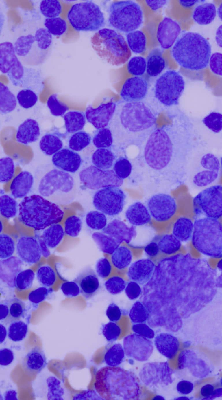
Sample images of Dr. Nebe's analyses
Which other technical aspects are crucial from your perspective?
The devil is in the detail. As already mentioned, the metachromatic staining method of the step-by-step May-Grünwald-Giemsa staining solution is unmatched to this day, especially due to the so-called Romanowski effect of this dye combination. Attempts to replace them with rapid-staining or single-step staining (e.g., Wright staining) have failed to convince experts. Light sources used to stimulate these colors – first sunlight reflectors, then incandescent bulbs, followed by halogen lamps (including thermal blue and gray filters to adjust and correct the color temperature) and, finally, light-emitting diodes of different color spectra – generate different spectra at the excitation wavelength and, thus, different absorption behavior of the complex dye reaction during the sophisticated MGG coloration.
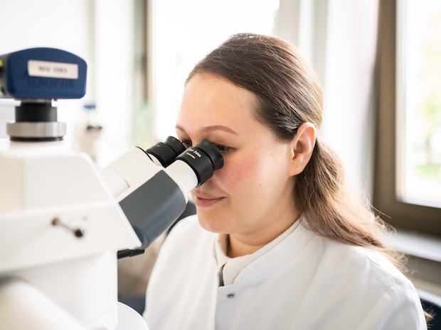
The selection of suitable LEDs has kept microscope manufacturers busy in the last few years. The development of optics since van Leeuwenhoek is limited in its resolution by the wavelength of light, as described by Abbe. Nevertheless, the quality of the lenses plays a major role (selecting the right glass and correcting the chromatic aberrations) and has been perfected in the lens systems of the plan apochromats. However, the image quality is also limited by the number of lenses required for this purpose (loss of contrast). The plan neofluars are a good compromise here. As agreed by cytology experts, the factors of overview, magnification, resolution and depth of field, reach their optimum with the 63x Plan-Apo lens for blood smears and 40x for bone marrow smears. Projecting this image on the camera chip with the correct magnification, thereby making optimum use of the resolution of the 4K chip by means of the camera tube, is the final contribution of the optical system to the perfect image. However, the contribution to the image quality of the condenser lens in the microscope is often forgotten (including cleaning it).
Could you also tell us something about your hematological samples?
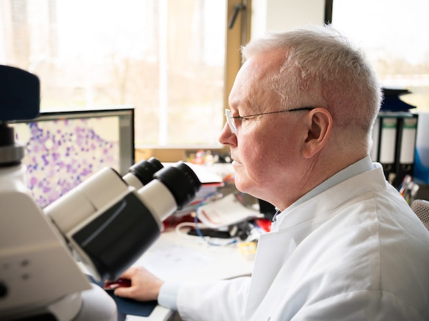
The evaluation of blood and bone marrow smears contributes significantly to the diagnosis of patients with hematological diseases because it integrates all cell lineages including their differentiation, maturation, activation and apoptosis. Pappenheim staining allows the cytological differentiation of healthy hematopoietic cells in reactive states on the one hand and, on the other, their deviation from normal differentiation in malignant transformation, e.g., in lymphoid neoplasms, myeloid dysplasias and, to a limited extent, in acute leukemias. What matters is the difference in size, nuclear/cytoplasmic ratio, chromatin structure (purple color shades), and cytoplasmic composition with very different granules (light and dark purple, reddish (eosin) or pinkish in monopoiesis) and its protein composition (shades of blue). They make the difference, as shown in an image of bone marrow from a patient with myelodysplasia (MDS), a pre-existing condition to acute myeloid leukemia (AML).
Where Pappenheim staining, which has been clinically validated for over 100 years (please rest here for a moment), reaches its limits, and new methods such as immunophenotyping (FACS), cytogenetics (FISH), and molecular genetics (PCR, NGS) come into play to complement but not replace morphology. However, until now, taken each of these more specific tests alone, they are decisive in less than 50% of the cases. By this means, morphology is the most integrative part in diagnosing hematological diseases.
How does your JENOPTIK GRYPHAX® ARKTUR camera help in hematopathology?
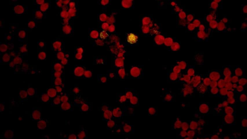
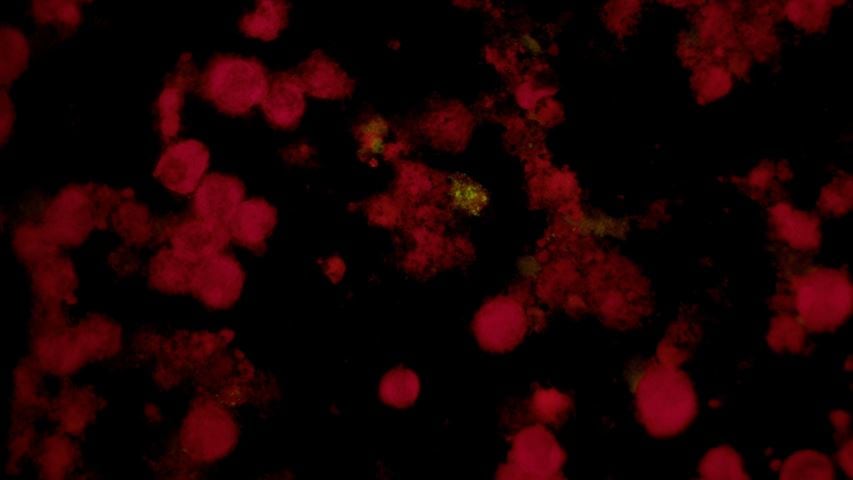
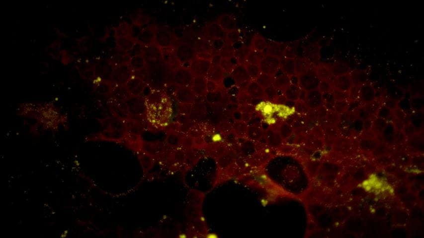
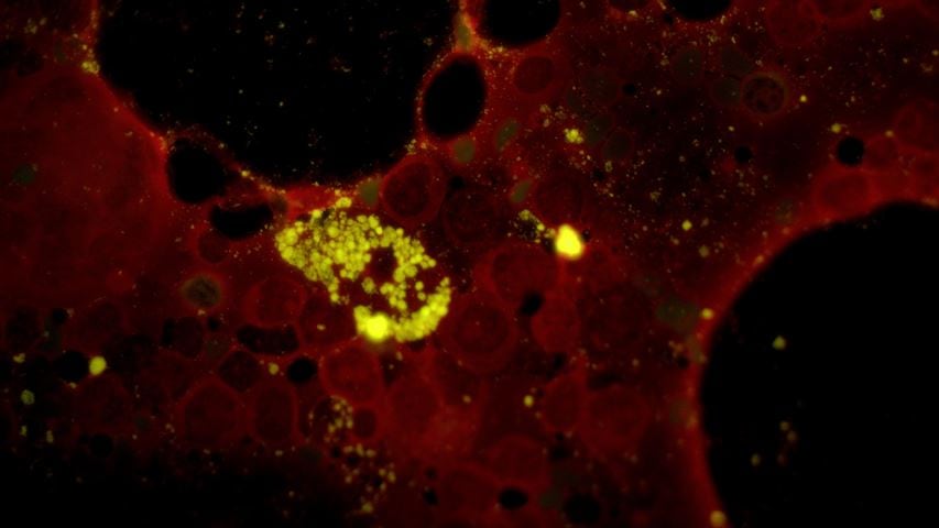
In addition to panoptic staining, fluorescence staining with antibodies is also used. Here, besides color reproduction, resolution and light sensitivity are very important. Digital fluorescence photography started with cooled black-and-white cameras. Subsequently brightnesses of separated color spectra of multi color imaging were converted into artificial colors by software and finally the images of various filter combinations were superimposed. Here, the JENOPTIK GRYPHAX® ARKTUR camera enables excellent reproduction in true color with short exposure times, shown here in an immunofluorescence image of Chlamydophila pneumoniae (FITC-labeled monoclonal antibody against Chlamydia LPS (green) and protein counterstain with Evans-Blue (red)) (please refer to slide images above). The 4K resolution (3840x2160) is the prerequisite for reproducing the higher image resolution in fluorescence microscopy. With 1920x1080, HDMI cannot adequately display the necessary details. In addition, to achieve color fastness, the image must be corrected in the camera and not on the computer. The chip should therefore guarantee good resolution, color fastness and contrast even in low light conditions over a wide dynamic range.
As regards the software, what do you appreciate most about your JENOPTIK GRYPHAX® ARKTUR camera?
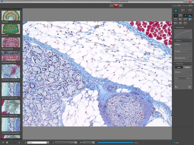
The software enables the user to directly control and adjust the camera for his needs. At the same time, the application is so intuitive and simple enough that you can concentrate on your work instead of devoting your valuable time just to technical aspects, which is certainly not common in this sector. This encompasses the time needed for correcting the white balance and brightness between the subsequent shots, including different dark and bright areas of the tissue and magnification of the object at different light intensity (color temperature). These tasks have been made easy by a special auto mode. Jenoptik has managed to find a good compromise here.
The further development of software is a continuous process involving customers, manufacturers and resellers. Here, ergonometric adjustments to the laboratory workflow have recently been achieved, making it easier for users to include microscopic images into their reports and support barcoded slides. In addition to ease of use, stability is also essential for daily routine, especially in patient care. The hardware is the one side, the software the other. The latter includes the "operating system" risk, which updates and changes continuously like a moving target for programmers and can cause problems as we all know. Therefore, I appreciate Jenoptik's product philosophy of self-sufficient software that provides all drivers and libraries by itself. Certainly a good decision, as shown by more than two years' experience. I also value good relationships with microscope dealers and laboratory software (LIS) suppliers for the important aspect of software integration, supported by Jenoptik's software development kit.
Products that might interest you further:


